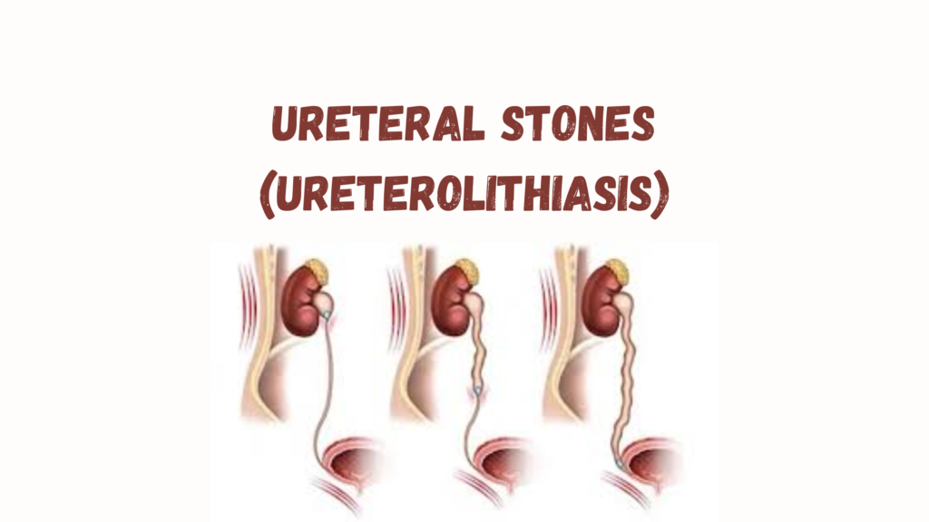
Ureteral Stones
Also called ureterolithiasis, these are kidney stones that have moved into the ureter, the narrow tube connecting the kidney to the bladder.
🔍 Pathophysiology
- Stones form in the kidney and may migrate down the ureter, causing pain, obstruction, or infection.
- Most stones are composed of:
- Calcium oxalate or phosphate (most common)
- Uric acid
- Struvite (infection-related)
- Cystine (rare, genetic)
⚠️ Symptoms
Classically known as renal colic:
- Sudden, severe flank pain radiating to the lower abdomen or groin
- Hematuria (blood in urine)
- Nausea and vomiting
- Urinary urgency or frequency (especially if the stone is near the bladder)
- Fever/chills (if infection is present – emergency)
📏 Stone Location Matters
| Location | Common Symptoms |
|---|---|
| Proximal ureter | Flank pain |
| Mid-ureter | Abdominal or back pain |
| Distal ureter (UVJ) | Groin pain, urinary symptoms |
🧪 Diagnosis
1. Imaging
- Non-contrast CT scan (CT KUB) – gold standard, detects nearly all types of stones.
- Ultrasound – useful in pregnant women and children, shows hydronephrosis.
- X-ray (KUB) – for radiopaque stones (e.g., calcium-containing).
- IVP or MR urogram – in selected cases.
2. Urinalysis
- Microscopic hematuria common
- Check for signs of infection (WBCs, nitrites)
3. Blood tests
- Renal function (creatinine, BUN)
- Electrolytes (especially calcium, uric acid)
- CBC if infection suspected
🩺 Treatment
Depends on:
- Stone size
- Location
- Symptoms
- Presence of infection or obstruction
🔹 Conservative Management (for small stones <5–6 mm)
- Hydration: Drink plenty of water.
- Pain relief: NSAIDs (e.g., ibuprofen), or opioids if severe.
- Alpha blockers (e.g., tamsulosin): Help relax ureter and pass stone.
✅ Up to 80% of small stones pass spontaneously.
🔹 Intervention Required When:
- Stone >6 mm (less likely to pass)
- Severe symptoms or uncontrolled pain
- Obstruction with infection (emergency!)
- Bilateral obstruction or obstruction in a single functioning kidney
- Persistent stone >4–6 weeks
Options:
- Ureteroscopy with laser lithotripsy
- Shock wave lithotripsy (SWL) – for certain stone sizes/locations
- Percutaneous nephrolithotomy (PCNL) – for large or complex stones
- Ureteral stent or nephrostomy tube for decompression if infection or obstruction
💡 Prevention
Based on stone type, but general advice includes:
- Increase fluid intake (goal: >2.5 L urine/day)
- Limit sodium and oxalate-rich foods
- Reduce animal protein intake
- Thiazide diuretics for recurrent calcium stones
- Allopurinol for uric acid stones