Pneumothorax

💨 Pneumothorax – Overview

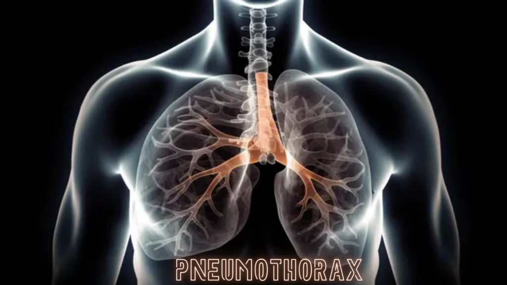
A pneumothorax is the presence of air in the pleural space (the space between the lung and the chest wall), which causes the lung to collapse partially or completely. This can lead to breathing difficulties and reduced oxygen levels.


⚙️ Types of Pneumothorax

TypeCause/Description
Spontaneous pneumothoraxOccurs without trauma; often in tall, thin young adults or people with lung disease (e.g., COPD)
Traumatic pneumothoraxDue to injury like rib fracture, stab wound, or chest surgery
Tension pneumothoraxLife-threatening; air enters pleural space and cannot escape, increasing pressure and compressing lung and heart

📋 Symptoms

  • Sudden onset sharp chest pain
  • Shortness of breath
  • Rapid breathing and heart rate
  • Decreased or absent breath sounds on affected side
  • In tension pneumothorax: low blood pressure, distended neck veins, tracheal deviation (emergency signs)

🩺 Diagnosis

  • Physical exam: decreased breath sounds, hyper-resonance on percussion on affected side
  • Chest X-ray: shows air in pleural space and lung collapse
  • Ultrasound: useful in emergency or bedside setting

💊 Treatment

SeverityTreatment
Small, uncomplicatedObservation and oxygen therapy; often resolves on its own
Larger or symptomaticNeedle aspiration or chest tube insertion to remove air
Tension pneumothoraxImmediate needle decompression followed by chest tube insertion (medical emergency)

⚠️ Complications

  • Respiratory failure
  • Recurrent pneumothorax
  • Infection if chest tube inserted

🛡️ Prevention

  • Avoid smoking (increases risk of spontaneous pneumothorax)
  • Careful monitoring of lung disease patients
  • Prompt treatment of chest trauma

Call Now Button