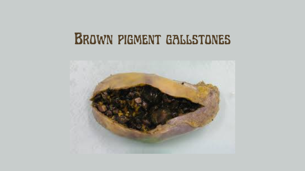
Brown pigment gallstones are a type of gallstone primarily composed of calcium bilirubinate, cholesterol, and fatty acids such as calcium palmitate and stearate. They are softer and greasier than black pigment stones and are predominantly found in the bile ducts rather than the gallbladder. These stones are commonly associated with biliary tract infections, biliary stasis, and parasitic infestations.(pmc.ncbi.nlm.nih.gov)
🧬 Pathogenesis
Brown pigment stones form when bacterial or parasitic infections in the biliary tract lead to the hydrolysis of conjugated bilirubin by bacterial β-glucuronidase. This process increases the concentration of unconjugated bilirubin, which then precipitates as calcium bilirubinate. Additionally, bacterial phospholipases hydrolyze biliary phospholipids, producing fatty acids that combine with calcium to form insoluble soaps, further contributing to stone formation. (pmc.ncbi.nlm.nih.gov)
⚠️ Risk Factors
- Biliary tract infections: Infections with enteric bacteria such as E. coli, Clostridium spp., and Bacteroides spp. (pmc.ncbi.nlm.nih.gov)
- Parasitic infestations: Infections with liver flukes like Clonorchis sinensis and Opisthorchis viverrini, especially prevalent in East Asia. (pmc.ncbi.nlm.nih.gov)
- Biliary stasis: Conditions leading to impaired bile flow, such as biliary strictures, choledochal cysts, and juxtapapillary duodenal diverticula. (pubmed.ncbi.nlm.nih.gov)
- Chronic pancreatitis: Inflammation of the pancreas affecting bile flow.
🩺 Clinical Features
Symptoms of brown pigment gallstones may include:
- Right upper abdominal pain: Often after meals.(health.com)
- Jaundice: Yellowing of the skin and eyes.
- Fever and chills: Indicating possible infection.(pmc.ncbi.nlm.nih.gov)
- Nausea and vomiting: Especially during or after meals.
These symptoms may suggest complications like cholangitis or biliary obstruction.(emedicine.medscape.com)
🧪 Diagnosis
Diagnostic approaches include:
- Ultrasound: To detect stones and assess bile duct dilation.
- CT or MRI scans: For detailed imaging of the biliary tract.
- Endoscopic retrograde cholangiopancreatography (ERCP): Allows for visualization and removal of stones from the bile ducts.
- Blood tests: To evaluate liver function and detect signs of infection or inflammation.
🛠️ Treatment Options
- Endoscopic procedures: ERCP is commonly used to remove stones from the bile ducts.
- Cholecystectomy: Surgical removal of the gallbladder may be considered if the gallbladder is involved.
- Antibiotics: To treat underlying infections.
- Parasitic treatment: Antiparasitic medications for infections like Clonorchis sinensis.(pmc.ncbi.nlm.nih.gov)
🌍 Epidemiology
Brown pigment gallstones are more prevalent in East Asia, particularly in regions where parasitic infections are common. In contrast, black pigment stones are more frequently encountered in Western countries. (pmc.ncbi.nlm.nih.gov, pmc.ncbi.nlm.nih.gov)